Proliferation assay - flow cytometry
Measuring stimulation-induced proliferation will give insight into the frequency of T cells that are responsive to a specific antigen of interest. This assay can then, for example, be used to assess modulation of this response by (immunomodulatory) compounds. Whether you are interested in modulation of allogeneic or antigen-specific reactions, or simply want to test the immunogenicity of your compound, our fully customizable flow cytometry-based proliferation assay will help you answer all of your questions.
If you want to know more about the functionality of T cells in response to your compound, this proliferation assay can easily be combined with measurements of cytokine production or transcription factor expression.
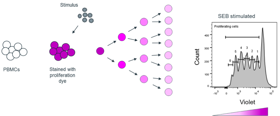
For our lymphocyte proliferation assay, we label the cells with a fluorescent proliferation dye. As the dye evenly dilutes out over both daughter cells upon cell division, each round of division can be tracked individually over time (see infographic). Because our assay is flow cytometry-based, the proliferation of a wide variety of individual cell types can be quantified simultaneously in the same sample.
Advantages of this proliferation assay
-
Precise monitoring of the number of cell divisions over a pre-determined period of time.
-
Easily combinable with other flow cytometry-based read-outs, such as cytokine production, cellular phenotyping and transcription factor expression.
- Specific cell subsets can be gated from the starting population upon read-out, i.e. the proliferation of individual subpopulations can be traced.
Also see our other proliferation assay by 3H thymidine incorporation.
Example of assessing proliferation via flow cytometry
Assessing T cell proliferation and transcription factor expression after cytomegalovirus (CMV) and tetanus toxoid (TT) peptide stimulation
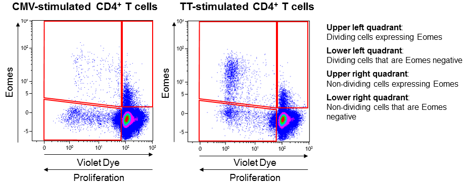
Violet stained PBMCs from a healthy donor were stimulated with either Cytomegalovirus (CMV) or Tetanus toxoid (TT). After 8 days, both antigens elicited a T cell response, which was measured by analyzing fluorescence in the violet spectrum. When examining expression of the transcription factor Eomes, it became clear that, while the rate of proliferation was comparable overall, stimulation with TT also induced marked upregulation of Eomes, while CMV stimulation did not. This indicates that different types of memory T cells respond to each of these antigens.
For more applications, please see the references below.
References
Guidelines for analysis of low-frequency antigen-specific T cell results: Dye-based proliferation assay vs 3 H-thymidine incorporation
Di Blasi D, Claessen I, Turksma AW, van Beek J, ten Brinke A.
J Immunol Methods 2020;487:112907. doi: 10.1016/j.jim.2020.112907.
Monitoring T-Cell Responses in Translational Studies: Optimization of Dye-Based Proliferation Assay for Evaluation of Antigen-Specific Responses
ten Brinke A, Marek-Trzonkowska N, Mansilla MJ, Turksma AW, Piekarska K, Iwaszkiewicz-Grześ D, Passerini L, Locafaro G, Puñet-Ortiz J, van Ham SM, Hernandez-Fuentes MP, Martínez-Cáceres EM, Gregori S.
Front Immunol 2017;8:1870. doi: 10.3389/fimmu.2017.01870.
Assay options
We offer proliferation analysis via flow cytometry in combination with several different standardized stimulation assays, but feel free to contact us for an assay customized to your needs.
Combine proliferation assessment via flow cytometry with
This flow cytometric approach is especially suited to combine with the following assays: